| <<< Previous | Index | Next >>> |
Case 12
Clinical and haematological data.
30-yr-old lady (MA).
Presented with renal failure, hypercalcemia and multiple osteolytic lesions.
Adenopathy, splenomegaly, no hepatomegaly. Anaemia. No paraprotein in the serum. Neutrophils precursors on the PB smear.
BM aspirate : dry tap.
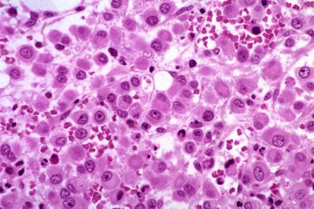
BM biopsy : diffuse infiltration by large cells characterized by eccentric nuclei, prominent nucleoli, abundant and dense cytoplasms without granules.
IHC : tumour cells positive for Melan A +, S100+, negative for keratin C11, CD138, Ig, CD45, B (CD20) - and T (CD3) - cell markers.
Interpretation and diagnosis: melanoma, metastatic.
Comments
BMB is more often positive for metastatic tumour than is aspirate, mainly because metastatic tumours induce fibrosis and or necrosis or may be too compact. In adults, breast carcinoma (female) and prostate and lung carcinoma (males) are the most frequent metastatic tumours involving the BM. In children, neuroblastoma is the most common solid tumour and has the highest frequency of BM involvement. In such situations, the panel of antibodies applied to BM sections is identical to that used in surgical pathology.
When the marrow is infiltrated by malignant cells, non-specific changes may be observed, including increased number of plasma cells, granulocytic and/or megakaryocytic hyperplasia and increased storage iron. A leucoerythroblastic reaction with circulating erythroblasts and immature granulocytes is a relatively common finding in the PB.
Malignant melanoma is found in the BM in approx. 5 % of patients with disseminated disease. Malignant melanoma should be suspected if metastatic tumour is composed of polygonal or spindle cells with prominent nucleoli. If melanin is present within the tumour cells, the diagnostic is relatively easy. However, not infrequently, metastatic melanoma is amelanotic and IHC mandatory for the diagnostic.
In the case presented here, the tumour cells are amelanotic and their morphological features can suggest a plasmablastic plasmocytoma or immunoblastic lymphoma and the correct diagnosis cannot be made without IHC.
Case 12. Melanoma, metastatic.
| <<< Previous | Index | Next >>> |
Copyright 2001, The Author(s) and/or The Publisher(s)
| Organisation: FORPATH asbl |
Coordination: Dr Bernard Van den Heule |
Host: Labo CMP |
