| <<< Previous | Index | Next >>> |
Case 10
Clinical and haematological data
31-yr-old female. Suffers from fatigue since 1 month. Presented with pancytopenia. No adenopathy, no hepatosplenomegaly. Hb 115 g/l, L 2 G/l, T 39 G/l
PB smear : rare blast cells.
BM histological examination
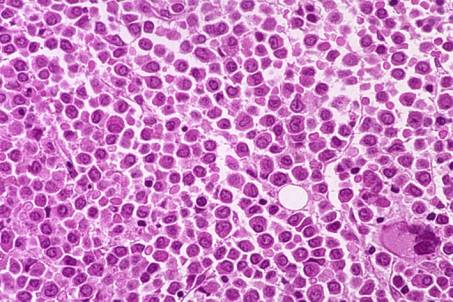
Increased cellularity, diffuse and monotonous cellular infiltration. The cells are medium-sized with somewhat eccentric rounded or lobulated nuclei, fine chromatin and dense non-granular cytoplasm.
Some residual islands of erythropoiesis and a few megakaryocytes are present. Focally, slight increase of the reticulin fibre network.
IHC: tumour cells positive for MPO and CD68/KP1 and negative for CD34,CD68/PGM1 Tdt, lymphoid markers and CD138.
Histologic interpretation and diagnosis
The differential diagnosis includes acute lymphoblastic leukaemia, multiple myeloma (some "plasma cell-like features"), Hairy Cell Disease and metastatic tumour. Positivity of the tumour cells for MPO allows a diagnosis of Acute Myeloid Leukaemia (AML), not further classified.
BM smears : blast cell population with granular cytoplasms and Auer rods and positive for Black Sudan. At flow cytometry: the blast cells co- express CD13 and CD33 and are positive for CD15 and CD65. Negativity for TdT and CD34.
Final diagnosis : AML M3 (FAB) according to the FAB system.
Comments
Histopathologists have to be familiarized with the morphological and IHC features of AL. BMB may viewed as complementary to the PB and BM smears for the diagnosis and follow-up of Acute Leukaemia (AL) (assessment of BM cellularity and residual haemopoiesis, quantity and rough characterization of the blasts). The trephine biopsy is necessary in cases showing dry tap at bone marrow aspiration or for classification of cases which lack circulating blasts.
Case 10. Acute Myeloid Leukaemia M3 (FAB)
| <<< Previous | Index | Next >>> |
Copyright 2001, The Author(s) and/or The Publisher(s)
| Organisation: FORPATH asbl |
Coordination: Dr Bernard Van den Heule |
Host: Labo CMP |
