CASE 6
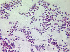
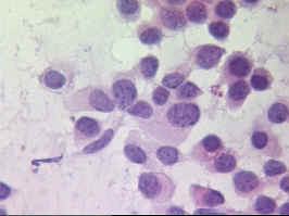
Clinical history and breast imaging: Screening mammography demonstrated a small stellate lesion at 12h in the left breast of a 71 years old patient. Ultrasound confirms a 1 cm large area of absorption at the same place. NCB and FNAC were guided by ultrasound.
Demonstrated slides: one representative smear.
| previous case | slide seminar | next page |
Copyright 1999, The Author(s) and/or The Publisher(s)
| Organisation: FORPATH asbl |
Coordination: Dr Bernard Van den Heule |
Host: Labo CMP |

